Make sure your sample meets these requirements before coming to image!
Read below for details…
When preparing samples for SIM, it is critical to use a #1.5 coverslip (~170 µm thickness). For best results, use “high-tolerance” (a.k.a. “high-precision”, “high-performance”) #1.5 coverslips with restricted thickness-related tolerance (± 5 µm). These are available from a number of vendors:
For live cell experiments, 35 mm glass-bottom dishes are also available in high-precision format:
Lab-Tek dishes may also be used on the microscope, but only the central four wells will be useable.
Ideally, coverslips should be cleaned via sonication or acid wash prior to use.
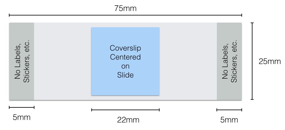
The travel range of the microscope stage is limited to the center 25×25 mm of the slide. Therefore, coverslips should be mounted in the center of a standard (25 x 75mm) slide, with no more than one coverslip per slide.
Fixed samples should be mounted in a high refractive index (> 1.45) mounting media with antifade. Frequently used mounting medias include:
Mounting media should NOT have any fluorophore (such as DAPI or Propidium Iodide). For nuclear counterstains, apply the counterstain and rinse away excess with multiple washes before mounting.
As a general rule, if the sample does not look good in widefield or confocal, SIM is not going to make it look better. Therefore, every attempt at sample optimization should be made before attempting SIM. Samples should be mounted such that the object of interest is as close to the coverslip as possible. Antibody concentration should be tested and labeling protocols should be optimized to reduce non-specific background from unbound fluorophore. Thick tissue should be cut to the smallest thickness consistent with experimental design to reduce background from out-of-focus fluorescence. For live cell experiments, imaging buffer should not include auto-fluorescent components such as phenol red.
Lastly, as a general rule, structured illumination works best when samples have have good inherent structure. Samples with diffuse fluorescence do not work as well.
A bright and photostable fluorophore is critical for good SIM reconstruction. For best results, chose fluorophores whose spectra match the lasers and filter spectra of the microscope. Use our filters spectra tool to compare the absorption and emission of your fluoropohore to the filters on our system.
If using fluorescent proteins, make every effort to update to the most recent proteins (i.e. don’t use mRFP, dsRed, or other dim and bleachy proteins). Protein performance will vary depending on sub-cellular environment, protein fusion, and imaging conditions, so testing multiple fluorophores may reveal a better match for your experiment.
For organic dyes, we have had good success with Alexa488, Alexa568, and Alexa647. (Alexa594 is acceptable, but 568 is preferable). Avoid older dyes such as FITC and TRITC.